Merke:
Wirkungsvoll die Out of Plane Punktion erlernen oder lehren
Mit welchen Übungen könntest du die Out of Plane-Punktion am schnellsten lernen und/oder lehren? Ich würde mit diesen drei aufeinander aufbauenden Punktionsübungen, die typisch für alltägliche klinische ultraschallgestützte Interventionen sind, anfangen.
Wenn du interessiert daran bist, deine knappe Zeit möglichst effektiv zum Erlernen der OOP-Punktionstechnik zu nutzen, höre dir den Podcast an oder besser noch, schaue dir das Video und Begleitmaterial auf der Webseite dazu an.
Inhaltsverzeichnis
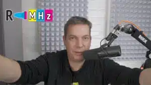
Podcast Out of Plane Punktion
Video(cast) Out of Plane Punktion
Der OOP-Podcast mit Videos und Bildern versehen
Auszug Transkript aus dem Podcast: „Ich bin neulich gefragt worden, sag mal Tim, wie geht denn ein Podcast über Ultraschall? Ist doch ein visuelles Verfahren und Podcast ist auditiv. Ja, da ist was Wahres dran, deswegen gibt es ja die Webseite.“
Beim Einsprechen des Podcasts habe ich mich gefilmt und dazu Material und Filme eingefügt, somit wird der auditive Podcast zum visuellen Videocast.
Learning Objectives der Out of Plane Punktionsübungen

Lies dir die drei Learning Objectives durch. Passend dazu habe ich die drei aus meiner Sicht wichtigsten Out of Plane-Punktionsübungen zusammengestellt, um möglichst schnell in der ultraschallgestützten Intervention Erfolge zu erzielen. Diese habe ich auf typisch anästhesiologische Prozeduren angewendet.
Die drei Übungen bauen aufeinander auf. Meine Empfehlung daher: arbeite sie in genau dieser Reihenfolge ab.
Erste Übung Out of Plane Punktion
Flacher Einstichwinkel für ultraschallgestützte Gefäßzugänge
Führe die Übung mit den typischen Kanülengrößen zur Gefäßpunktion durch (14-20G). Benutzt du andere Kanülen, weil du klinisch z. B. in der Kinderanästhesie arbeitest, verwende die entsprechend kleineren Kanülen.


Trotz Facettenschliff kein Doppelpunkt, wenn die Öffnung zur Sonde gerichtet ist.
Bei einem flachem Einstichwinkel wirst du KEINEN Doppelpunkt der Kanülenspitze mit Fazettenschliff sehen. Somit fällt ein wichtiges Kriterium zur sicheren Detektion des Kanülenendes weg.
Mit zunehmender Tiefe und steilerem Winkel ist der typische Doppelpunkt einer Facettenschliffkanüle in der Out of Plane-Punktionstechnik erkennbar (nächste Abbildung).

Schiebe die Kanüle in der Out of Plane-Technik bewusst über die Ebene hinaus. Beobachte die unterschiedlich erscheinenden Schatten des Schaftes, abhängig vom Punktionswinkel, Kanülendesign oder Darstellungstechnologie.

Zweite Übung Out of Plane Punktion
Steiler Einstichwinkel und etwas mehr Ultraschallphysik: die Schichtdicke
Lerne den Schichtdickenartefakt (slice thickness artifact) in der OOP-Technik bewusst zu nutzen, indem du mit steilem Winkel kurz vor der Sonde einstichst. Der steile Einstichwinkel ist hilfreich bei single-shot-Blockaden in der Regionalanästhesie.


Blogposts zur Schichtdicke auf Radiomegahertz
Die Bedeutung der Schichtdicke für die ultraschallgestützte Intervention wurde auf Radiomegahertz bereits eingehend behandelt. Klicke auf die Abbildungen, um auf die entsprechenden Beiträge zu gelangen, wenn du ein kurzes UpDate zur Schichtdicke haben möchtest.


Out of Plane-Punktionsübung mit steilem Einstichwinkel, sehr kleiner Kanüle (27G) mit der Kanülenöffnung mit Fazettenschliff zur Sonde gerichtet. Die Öffnung ist bei ruhiger Sondenhaltung und Kanülenführung eindeutig zu identifzieren.

Dritte Übung Out of Plane Punktion
Statische Sonde, flache und steile Einstichwinkel, Kanülenmanöver
Du lernst anhand des 4-Ecken-Out of Plane-Spiels, wann Winkelkorrekturen in größeren und kleineren Dimensionen durchgeführt werden (siehe Abbildung). Ziehe die Kanüle immer erst zur Oberfläche zurück (Hautniveau), bevor du die Kanülenmanöver durchführst.


Welches Punktionsphantom solltest du verwenden?
Ich plädiere für ein Phantom aus Fleisch. Der Grund dafür ist, dass Punktionstechniken klassischerweise mit Injektionen verbunden werden. Sofern keine kommerziell erhältlichen Phantome, die speziell für Injektionen entwickelt wurden, vorhanden sind, ist Fleisch eine sehr preiswerte Alternative. Sonoanatomisch betrachtet bieten die Faszien und Sehnen in einem größerem Stück Schweinefleisch ideale Punktionsziele. Denke dabei zum Beispiel an das Erüben der Hydrodissektionstechnik (nicht im Podcast besprochen). Geflügelfleisch ist gegenüber Rind und Schwein homogener und daher nicht nur aus Infektionsgründen weniger geeignet (s. unten).
Hygienische Aspekte zu Fleischphantomen
Die Ultraschallsysteme werden an Patient*innen eingesetzt. Eine vorausgehende und abschliessende Desinfektion ist selbstverständlich. Ein Sondenüberzug und Handschuhe beugen Kontaminationen vor. Die zur Übung benötigten Kanülen sollten über die üblichen Behälter entsorgt werden, um Stichkontaminationen zu vermeiden.
Mögliche sonografische Probleme von Fleischphantomen
Ist ein Fleischphantom immer gut zu sonografieren? Nein, denn häufig sind Areale zu detektieren, die von Ultraschallwellen nicht durchdrungen werden.
Häufig ist die Ursache, dass das Fleisch tiefgefroren wurde. Auch sind Lufteinschlüsse möglich. Du kannst diesen Problemen begegnen, indem du das Fleisch etwas auftauen lässt, was auch für die Schallweiterleitung günstig ist, oder in ein Wasserbad unter Bewegung einlegst.
Weil das alle keine Garantien für brilliante Ultraschallbedingungen sind, würde ich ein Phantom auf Reserve kaufen.

Punktionsübungen an Körperspendern in der Anatomie kombieren die Möglichkeit zugleich die menschliche Anatomie und Injektionstechniken zu erlernen. Wenn du bereits über gute Erfahrungen mit der in plane Punktion besitzt, könntest du du hier die out of plane Punktion trainieren. ErfahrungenSiehe Beitrag Sonoanatomie und Interventionen am Körperspender.

Gelatine ist für diese Punktionsübungen aufgrund der zu hohen Echogenität der Kanülen in dem nicht reflektierenden Medium weniger geeignet als Fleischphantome. Beitrag Shooting with Sound – Ballistic Gel.

Zusammenfassung
Die Entscheidung für drei Punktionsübungen sind bewusst so ausgefallen. Sie decken in der ersten Übung die Anlage peripher-venöser Verweilkanülen, A. radialis-Punktionen oder ZVK-Anlagen ab. Es wird der Unterschied zur single-shot-Blockade in der Regionalanästhesie in der zweiten Übung herausgearbeitet. Du lernst, wann du ein slide-down-Manöver durchführst und wann die Ultraschallsonde nahezu statisch gehalten wird. Hervorgehoben wird, dass das Ultraschallbild aus einem Volumen generiert wird und du die Effekte des Schichtdicken–Artefakt gezielt nutzen kannst. Übung drei verdeutlicht das aus meiner Sicht hervorragend. Alle drei Out of Plane-Punktionstechniken sind auf typisch anästhesiologische Prozeduren ausgelegt. Bespielhaft würden Interventionalisten der Inneren Medizin für eine Nierenbiopsie andere Übungen bevorzugen.
Merke: „Es gibt nichts Gutes, außer man tut es.“ Also ran an die Buletten, will sagen, an die Punktionsübungen.
Weitere Möglichkeiten der Punktionsübungen
Punktionsübungen im Kurs sinnvoll und notwendig, aber nicht ausreichend.
In einem Ultraschallkurs werden ähnliche Aufgaben praktisch umgesetzt und erarbeitet und sind fester Bestandteil in den Curricula. Dennoch reicht die im Kurs zur Verfügung stehende Zeit nicht aus, um mit profunder Fertigkeit den Kurs abschließen zu können. Aus meiner Sicht könnte man einen gesamten Tag nur für Punktionsübungen verwenden, um signifikante Erfolge in der klinischen ultraschallgestützten Intervention erfahren zu können.
Solange es keinen Kurs ausschließlich für Punktionsübungen gibt, ist Eigenarbeit erforderlich. Mit dem Podcast und dem Blogpost hast du nun eine Starthilfe. Und ergänzende Anregungen, wie du die beschriebenen Übungen erweitern könntest. Stelle dir auch selbst Übungen vor dem Hintergrund deiner klinischen Tätigkeit vor. Wichtig ist nur, dass du in die Umsetzung gehst und dir ein- zweimal ausreichend Zeit nimmst, um die manuellen Fertigkeiten zu erlangen. Dann kannst du klinisch weitermachen und die Techniken verfeinern.
Vorschläge
- Beobachte Niedrigvolumina-Injektionen vor, innerhalb und hinter dem Schichtdickevolumen.
- Suche dir Faszien und übe die Hydrodissektion, schaue dir dabei die Kanülenöffung an und vergleiche die OOP- mit der IP-Technik.
- Vergleiche Fazettenschliff-Kanülen mit anderen (Tuohy oder reguläre Spitze) und entscheide dich aufgrund sonografisch erhobener Erfahrungen.
- Filme dich während der Prozedur und analysiere deine Sonden- und Kanülenmanöver.
- Lerne etwas über die tilt-down-Technik und diskutiere mit Kolleg*innen darüber.
- Verändere den Blick auf die Sonde: schmale oder breite Seite und beobachte mögliche Abweichungen des Kanülenvorschubs.
- Verwende Sehnen, Drainagen oder wassergefüllte Schlangenluftballons als Punktionsziel.
- Punktiere auf Zeit im Wettbewerb mit Kolleg*innen, evaluiere dabei die Punktionsgenauigkeit gegenüber einem Zeitgewinn.
- Führe die out of plane Punktion oder Injektion lateral und medial deines Zieles aus.
- und und und
Feedback zum Beitrag
Hat die der Beitrag gefallen? Wenn ja, freue ich mich über eine Rückmeldung. Wenn nicht, schreibe mir doch warum. Email an: support@radiomegahertz.de.
Referenzen und Links
Liu, L.; Tan, Y.; Li, S.; Tian, J. “Modified Dynamic Needle Tip Positioning” Short-Axis, Out-of-Plane, Ultrasound-Guided Radial Artery Cannulation in Neonates: A Randomized Controlled Trial. Anesth Analg 2019, 129, 178–183. doi:10.1213/ANE.0000000000003445
Takeshita, J.; Nakayama, Y.; Tachibana, K.; Nakajima, Y.; Shime, N. Ultrasound-guided short-axis out-of-plane approach with or without dynamic needle tip positioning for arterial line insertion in children: A systematic review with network meta-analysis. Anaesth Crit Care Pain Med 2023, 42, 101206.
Wu, G.; Chen, C.; Gu, X.; Yao, Y.; Yuan, D.; Lv, J.; Zhao, B.; Wang, Q. Ultrasound-Guided Dynamic Needle-Tip Positioning Method Is Superior to Conventional Palpation and Ultrasound Method in Arterial Catheterization. J Clin Med 2022, 11, 6539. doi:10.3390/jcm11216539
Liang, Y.; Liu, P.; Wei, C.; Li, W.; Li, C.; Lai, T.; Peng, S.; Xu, J.; Zhang, H.; Li, P.; Li, S. Ultrasound-guided modified dynamic needle tip positioning technique for distal radial artery catheterization: A randomized controlled trial. J Vasc Access 2024, 11297298241270537. doi:10.1177/11297298241270537
Clemmesen, L.; Knudsen, L.; Sloth, E.; Bendtsen, T. Dynamic needle tip positioning – ultrasound guidance for peripheral vascular access. A randomized, controlled and blinded study in phantoms performed by ultrasound novices. Ultraschall Med 2012, 33, E321–5. doi:10.1055/s-0032-1312824
English version of this blogpost
Note
This translation from German into English was carried out using ChatGPT 4o (accessed on 17 October 2024, two days after the release of this blogpost).
If an ultrasound image is not good, the rule usually applies: Don’t blame the machine or the patient. You are the cause of the mediocre image.
However, in this case of the translation, the rule is: Blame the machine, it wasn’t me.
Effectively learn or teach the out-of-plane puncture
What exercises could help you learn and/or teach the out-of-plane puncture most quickly? I would start with these three progressive puncture exercises, which are typical for everyday clinical ultrasound-guided interventions.
If you are interested in utilizing your limited time as effectively as possible to learn the OOP puncture technique, listen to the podcast or, better yet, watch the video and accompanying materials on the website.
Video(cast) Out of Plane Punktion
The OOP podcast supplemented with videos and images.
Excerpt from the podcast transcript:
„I was recently asked, ‚Hey Tim, how does a podcast about ultrasound work? It’s a visual procedure, and a podcast is auditory.‘ Yes, there’s some truth to that, which is why there’s a website.“
While recording the podcast, I filmed myself and included materials and videos, turning the auditory podcast into a visual videocast.

Read through the three learning objectives. In line with them, I’ve put together what I consider to be the three most important out-of-plane puncture exercises to achieve success in ultrasound-guided interventions as quickly as possible. I’ve applied these to procedures typically performed in anesthesiology.
The three exercises build on each other. My recommendation, therefore, is to work through them in exactly this order.
First Exercise: Out-of-Plane Puncture
Shallow puncture angle for ultrasound-guided vascular access
Perform the exercise using the typical needle sizes for vascular puncture (14-20G). If you use different needles because you work clinically in pediatric anesthesia, use the appropriately smaller needles.


Despite the bevel tip, there is no double point when the opening is directed towards the probe.
With a shallow puncture angle, you will NOT see a double point of the bevel tip needle. Thus, an important criterion for the safe detection of the needle tip is lost.
With increasing depth and a steeper angle, the typical double point of a bevel-tip needle becomes visible in the out-of-plane puncture technique (next image).

Deliberately advance the needle beyond the plane in the out-of-plane technique. Observe the differently appearing shadows of the shaft, depending on the puncture angle, needle design, or imaging technology.

Second Exercise: Out-of-Plane Puncture
Steeper puncture angle and a bit more ultrasound physics: the slice thickness
Learn to consciously utilize the slice thickness artifact in the out-of-plane technique by puncturing at a steep angle just before the probe. The steep puncture angle is helpful for single-shot blocks in regional anesthesia.


Blog posts on slice thickness at Radiomegahertz.de
The significance of slice thickness for ultrasound-guided intervention has already been thoroughly discussed on Radiomegahertz. Click on the images to access the relevant posts if you would like a quick update on slice thickness.
Out-of-plane puncture exercise with a steep puncture angle, using a very small needle (27G) with the bevel opening directed towards the probe. The opening can be clearly identified with stable probe positioning and needle guidance.

Third Exercise: Out-of-Plane Puncture
Static probe, shallow and steep puncture angles, needle maneuvers
You will learn through the Four Corners Out-of-Plane game when to make angle corrections in larger and smaller dimensions (see image). Always retract the needle back to the surface (skin level) before performing the needle maneuvers.


Which puncture phantom should you use?
I advocate for a meat phantom. The reason for this is that puncture techniques are traditionally associated with injections. If commercially available phantoms specifically designed for injections are not available, meat is a very cost-effective alternative. From a sonoanatomical perspective, the fasciae and tendons in a larger piece of pork provide ideal puncture targets. For instance, consider practicing the hydrodissection technique (not discussed in the podcast). Poultry is more homogeneous compared to beef and pork and is therefore less suitable, not only for infection reasons (see below).
Hygienic aspects of meat phantoms
The ultrasound systems are used on patients. Preliminary and final disinfection is, of course, required. A probe cover and gloves prevent contamination. The needles used for practice should be disposed of in the usual containers to avoid needlestick contamination.
Possible sonographic issues with meat phantoms
Is a meat phantom always easy to scan sonographically?
No, because areas are often detected that cannot be penetrated by ultrasound waves.
The cause is often that the meat has been frozen. Air pockets are also possible. You can address these problems by allowing the meat to thaw slightly, which also improves sound transmission, or by placing it in a water bath with movement.
Since none of these methods guarantee optimal ultrasound conditions, I would recommend buying a backup phantom.

Puncture exercises on body donors in anatomy combine the opportunity to simultaneously learn human anatomy and injection techniques. If you already have good experience with in-plane punctures, you could train out-of-plane punctures here. See the post on sonoanatomy and interventions on body donors.

Gelatin is less suitable for these puncture exercises than meat phantoms due to the high echogenicity of the needles in the non-reflective medium. See the post „Shooting with Sound – Ballistic Gel.„

In summary
The decision to include three puncture exercises was made deliberately. The first exercise covers the placement of peripheral venous catheters, radial artery punctures, or central venous catheter insertions. The difference from single-shot blockade in regional anesthesia is highlighted in the second exercise. You will learn when to perform a slide-down maneuver and when to hold the ultrasound probe almost static. It is emphasized that the ultrasound image is generated from a volume, and you can intentionally use the effects of the slice-thickness artifact. Exercise three illustrates this excellently from my point of view. All three out-of-plane puncture techniques are tailored to typical anesthesiology procedures. By contrast, interventionalists in internal medicine, for example, might prefer different exercises for a kidney biopsy.
Remember: „There is nothing good unless you do it.“ So get to it—time to tackle the puncture exercises.
Additional options for puncture exercises
Puncture exercises in the course are useful and necessary, but not sufficient enough for clinical routine.
In an ultrasound course, similar tasks are implemented and worked on practically, and they are a fixed part of the curricula. However, the time available in the course is not sufficient to complete it with a solid skill set. In my view, an entire day could be dedicated solely to puncture exercises to achieve significant success in clinical ultrasound-guided interventions.
As long as there is no course exclusively for puncture exercises, self-study is required. With the podcast and blog post, you now have a starting point. Additionally, here are some suggestions on how you could expand upon the exercises described. Also, envision exercises in the context of your clinical work. The important thing is to take action and set aside time once or twice to acquire the manual skills. Then you can continue clinically and refine the techniques.
Suggestions
- Observe low-volume injections before, within, and behind the slice thickness volume.
- Identify fascial planes and practice hydrodissection, paying attention to the needle opening and comparing out-of-plane (OOP) techniques with in-plane (IP) techniques.
- Compare bevel-tip needles with others (e.g., Tuohy or regular tips) and make decisions based on sonographically obtained experiences.
- Film yourself during the procedure and analyze your probe and needle maneuvers.
- Learn about the tilt-down technique and discuss it with colleagues.
- Change your perspective on the probe: narrow or wide side, and observe possible deviations in needle advancement.
- Use tendons, drains, or water-filled balloon animals as puncture targets.
- Compete with colleagues to puncture within a set time, evaluating puncture accuracy against time efficiency.
- Perform out-of-plane punctures or injections laterally and medially of your target.
- And so on.